
Nuclear Medicine
Diagnostic Imaging Services
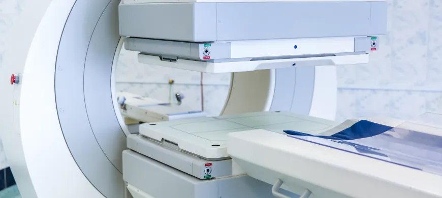
Nuclear medicine uses very small, safe amounts of radioactive materials, called radiopharmaceuticals, to image specific organs and functions within the body.
A gamma camera detects the radiopharmaceuticals and
produces 3D images for evaluation by a radiologist or
cardiologist.
If you are pregnant, please discuss with your physician prior to scheduling your test and notify the schedulers when making your appointment, as well as the technician at the imaging center.
Maximum weight capacity for our nuclear medicine equipment: 400 lbs.

Bone Scan
A bone scan is used to determine if you have abnormalities within your bones such as small stress fractures, cancer, tumors, infection or to determine the cause of unexplained pain.
Preparation
If you are pregnant or think you might be, please inform the technologist.
It is important that you drink a lot of fluids prior to the test. This will improve the quality of your images. You may take your medications and eat before your test.
What to expect during the test
Upon arrival, we will review your medical history. You will be given an injection of a small amount of radioactive tracer. This is not a drug or dye. After the injection, you will be permitted to leave for 2 hours during which time we ask that you drink a minimum of 32 ounces of fluid. Upon your return you will be placed on a narrow imaging bed to allow the camera to pass over and below you taking images of your bones for approximately 20-40 minutes. You will be asked to remain still during the pictures. In some cases, your doctor might order a three-phase bone scan, which includes a series of images taken at different times. A number of images are taken as the tracer is injected, then again shortly after the injection and two to four hours later.
Liver/Spleen Scan
A liver scan may be performed to screen for diseases such as cancer, hepatitis, or cirrhosis. Lesions such as tumors, abscesses, or cysts of the liver or spleen may be seen on a liver scan. A liver scan may be performed to assess the condition of the liver and/or spleen after trauma to the abdomen or when there is unexplained pain in the right upper quadrant of the abdomen. Enlargement of the liver or spleen may be seen on a liver scan.
Preparation
If you are pregnant or think you might be, please inform the technologist, otherwise, no prep.
What to Expect During the Test
You will be given an injection of a small amount of a radioactive tracer. This is not a drug or dye. Approximately 15 minutes after the injection, you will lie down on a narrow, padded table. A special camera will be lowered close to your body. Images will be taken for approximately 20-30 minutes.
Hepatobiliary Scan
A hepatobiliary scan is used to determine if your gallbladder is functioning properly in the absence of gallstones. Additionally, it assists in determining if abdominal pain or nausea is related to gall bladder disease.
Preparation
If you are pregnant or think you might be, please inform the technologist. Do not eat or drink anything 4 hours prior to the test.
What to expect during the test
Upon arrival, we will review your medical history. A small I.V. will be inserted into your arm through which you will be given an injection of a small amount of a radioactive tracer. After the injection, you will be placed on a narrow imaging bed to allow pictures to be taken of your gallbladder. You will be asked to remain still as images are taken every 15 minutes. After 1 hour, you will be given medication through your I.V. that should make your gallbladder contract, with additional images taken 15 and 30 minutes later. This medication may make you feel warm or nauseated for a few minutes. This sensation will pass quickly, typically within a few minutes.
Renal (Kidney) Scan
A renal or kidney scan is used to determine how well blood is flowing to your kidney and how well the kidneys are functioning. In some cases, this test can be used to determine if poor blood flow to the kidneys is a result of prolonged high blood pressure.
Preparation
- If you are pregnant or think you might be, please inform the technologist.
- No food 5 hours prior to the test.
- You must drink a minimum of 32 ounces of fluid one hour before your appointment.
- Diuretics, ACE Inhibitors and Angiotensin II Blockers must be stopped a minimum of 48 hours prior to exam.
- Beta Blockers, Calcium Channel Blockers and Alpha Blockers can be continued.
- Contact your physician or pharmacist to ask if your medications fall in one of these categories.
What To Expect During the Test
Upon arrival, we will review your medical history. A small I.V. will be started in your arm. In some cases you will be given a pill and have your blood pressure monitored for one hour. You will be placed on a narrow imaging bed to allow pictures to be taken of your kidneys. You will be given an injection of a small amount of radioactive tracer through your I.V. You will be asked to remain still as pictures are taken for 30 minutes. After 30 minutes, you will be asked to empty your bladder and return for a one-minute picture.